Ralph Perry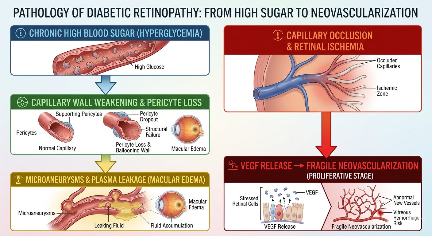
Diabetes has reached epidemic proportions globally, and among its most severe complications is its impact on ocular microvasculature. Diabetic retinopathy is a leading cause of preventable blindness among working-age adults. The condition develops quietly, often progressing for months or years without causing noticeable blurriness, pain, or discomfort.
Jump To
- How Diabetes Affects the Retinal Microvasculature?
- The Stages of Diabetic Eye Disease
- Stage
- Pathological Features
- Common Symptoms
- Clinical Recommendation
- High-Tech Diagnostic Modalities for Retinal Health
- Modern Treatment Interventions for Retinal Protection
- Protect Your Vision at Infinite Vision Care
For anyone living with Type 1 or Type 2 diabetes, partnering with a fellowship-trained Ophthalmologist in Mira Road or visiting an advanced eye clinic in Mira Road for regular retinal screenings is critical to safeguarding long-term vision.
How Diabetes Affects the Retinal Microvasculature?
The retina is the light-sensitive neural layer at the back of the eye, densely packed with microscopic capillaries that supply oxygen and nutrients to photoreceptor cells. Chronic hyperglycemia (elevated blood glucose) damages the delicate endothelial walls of these tiny vessels over time.
As the capillary walls weaken, fluid and fatty lipids leak into the retinal tissue. When this leakage occurs in the macula—the central zone responsible for sharp, reading vision—it causes Diabetic Macular Edema (DME), leading to wavy distortions and central blurring.
The Stages of Diabetic Eye Disease
Understanding the clinical progression of diabetic retinopathy helps patients recognize why routine screening is essential even when vision feels normal.
Stage |
Pathological Features |
Common Symptoms |
Clinical Recommendation |
|
Mild NPDR (Non-Proliferative) |
Presence of microaneurysms (tiny balloon-like swellings in retinal blood vessels). |
Asymptomatic (Vision is typically 6/6). |
Strict glycemic control; annual dilated fundus exam. |
|
Moderate to Severe NPDR |
Blocked vessels, hemorrhages, cotton-wool spots, and retinal ischemia. |
Mild fluctuations in vision; slight contrast reduction. |
Monitoring every 3 to 6 months; OCT scanning. |
|
PDR (Proliferative Retinopathy) |
Severe oxygen deprivation triggers abnormal, fragile new blood vessels (neovascularization). |
Sudden showers of floaters, flashes, cobweb vision, severe sudden vision loss. |
Urgent Pan-Retinal Photocoagulation (Laser) or Anti-VEGF therapy. |
|
Diabetic Macular Edema (DME) |
Fluid accumulation and swelling in the central macula (can occur at any stage). |
Distorted straight lines, washed-out colors, difficulty reading small print. |
Intravitreal Anti-VEGF / Steroid micro-injections. |
High-Tech Diagnostic Modalities for Retinal Health
A standard vision chart check cannot evaluate the health of the retina. An advanced eye hospital in Mira Road utilizes specialized imaging platforms:
- Dilated Indirect Ophthalmoscopy: Wide-field illumination of the entire retinal periphery to detect peripheral hemorrhages or early neovascularization.
- Optical Coherence Tomography (OCT): A non-invasive cross-sectional scan that measures retinal thickness down to single-micron resolution, diagnosing subclinical macular swelling before it causes visual symptoms.
- Fundus Fluorescein Angiography (FFA): A specialized dye test that maps retinal blood flow, revealing capillary non-perfusion, leakage points, and abnormal vascular branching.
Modern Treatment Interventions for Retinal Protection
Early detection allows retinal specialists to halt disease progression and protect functional vision through minimally invasive, targeted therapies:
- Intravitreal Anti-VEGF Injections: Medications that block Vascular Endothelial Growth Factor (VEGF), the chemical signal that drives abnormal blood vessel growth and vascular leakage in the macula. Administered under local drop anesthesia, these quick injections reduce macular swelling and restore visual clarity.
- Pan-Retinal Photocoagulation (PRP Laser): Precision thermal laser spots applied to oxygen-starved peripheral retinal areas, shrinking abnormal blood vessels and preventing catastrophic vitreous hemorrhage or tractional retinal detachment.
- Focal / Grid Laser Photocoagulation: Seals localized leaking microaneurysms around the macula to stabilize central vision.
Protect Your Vision at Infinite Vision Care
Managing diabetic eye health requires meticulous diagnostic capability and super-specialized clinical care.
At Infinite Vision Care and Laser Centre, Dr. Shaila Rajkumar Patel provides comprehensive diagnostic and therapeutic eye care:
- Fellowship-Trained Precision: Extensive expertise in anterior and posterior segment evaluations, corneal mapping, and retinal monitoring.
- Comprehensive Care Pathway: Advanced in-house imaging (OCT, digital fundus evaluation) to detect early microvascular damage.
- NABH-Accredited Excellence: Providing a safe, patient-centric clinical environment with personalized management protocols.
If you or a loved one have diabetes, do not wait for vision to blur. Schedule a comprehensive dilated retinal evaluation with an experienced eye specialist in Mira Road or a leading Ophthalmologist in Kandivali at Infinite Vision Care today.